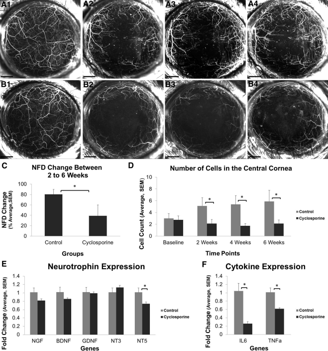
Figure 3.
Effect of CsA on nerve regeneration after lamellar corneal surgery. (A1–B4) Serial in vivo maximum intensity projection images of fluorescent nerves and inflammatory cells in a thy1-YFP mouse cornea before surgery (A1, B1) and 2 weeks (A2, B2), 4 weeks (A3, B3), and 6 weeks (A4, B4) after surgery. Eyes were treated with artificial tears (A1–A4) or CsA (B1–B4) twice daily for 6 weeks. The NFD growth between weeks 2 and 6 (C) as well as the number of YFP-positive cells in the central 2-mm cornea (D) are significantly lower in the CsA group. Neurotrophin expression (E) and inflammatory cytokine expression (F) at 6 weeks. Scale bar, 500 μm; *P < 0.05.

