Figure 4.
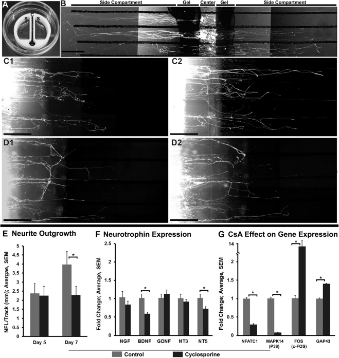
Effect of CsA on neurite outgrowth in compartmental cultures of dissociated trigeminal ganglion cells. (A) A device (Teflon) used for compartmental cultures of trigeminal ganglion neurons. The central compartment, which contains trypan blue dye, is isolated from side compartments containing culture media. Note the absence of leakage of the dye from the central compartment to the side compartments. (B) Fluorescent image showing neurites in the growth media. The cell bodies are isolated in the central compartment and the neurites extend into the side compartment parallel to the tracks. (C1–D2) Comparison of outgrowth between neurites exposed to vehicle (C1, C2) or CsA (D1, D2) for 24 hours. Neurite outgrowth was assessed on day 5 (C1, D1) before, and day 7 (C2, D2) after vehicle or CsA treatment. (E) Quantification of neurite outgrowth. The bars show the NFL per track before (day 5) and after (day 7) vehicle or CsA treatment. (F) Real-time qPCR analysis at day 7 shows that BDNF and NT5 gene expressions are significantly less after CsA treatment compared with vehicle treatment. (G) Real-time qPCR analysis of expression of genes that are specific to CsA mechanism of action (NFATC1, MAPK14, and FOS). Graph shows fold increase or decrease in gene expression in CsA-exposed neurons compared with gene expression in dissociated trigeminal neurons (control). CsA exposure significantly decreases the expression of NFATC1 and MAPK14 (P38) but significantly increases the expression of FOS (c-fos) and GAP43. *P < 0.05. Scale bar, 500 μm.

