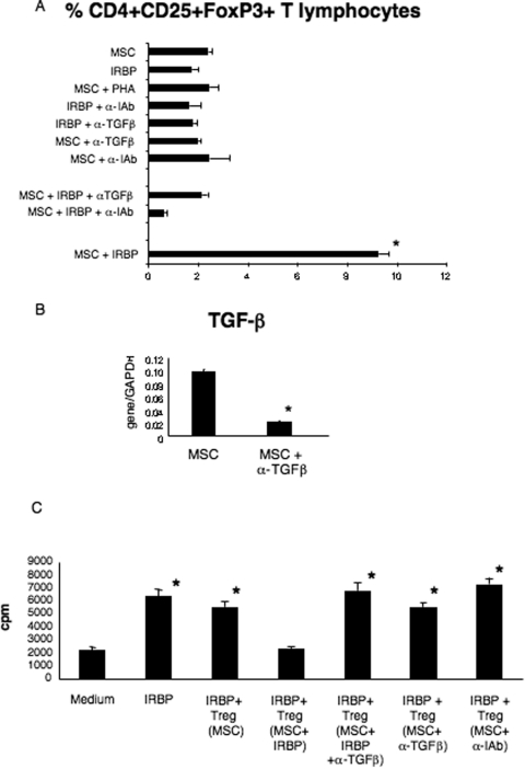
Figure 4.
Effect of α-MHC class II or α-TGFβ antibodies on in vitro conversion of CD4+ T lymphocytes into functionally active antigen-specific Treg. (A) Percentage of CD4+CD25+FoxP3+ T lymphocytes isolated from the top portion of transwell experiments in which splenocytes from naive mice were seeded. *P < 0.001 comparing the percentage of Treg generated in the presence of MSCs and peptide (MSC+IRBP) with all the other conditions. **P < 0.001 comparing wells in which Treg were generated in the presence of MSC, IRBP peptide, and α-IAb blocking antibodies (MSC+IRBP+α-IAb) with all the other conditions. (B) TGFβ expression by MSCs isolated from transwell plates in the absence (MSC) or presence of α-TGFβ blocking antibodies (MSC+α-TGFβ). Shown are the compiled results of three independent experiments. *P < 0.001 comparing the two different conditions. (C) Proliferation of T lymphocytes isolated from uveitic mice to the immunizing antigen in the presence or absence of CD4+CD25+FoxP3+ T lymphocytes (Treg) isolated from the transwell experiments. Shown are compiled results of three independent experiments. *P < 0.001 comparing the proliferation observed in any experimental condition with the baseline value (medium). Bars indicate mean + SD.

