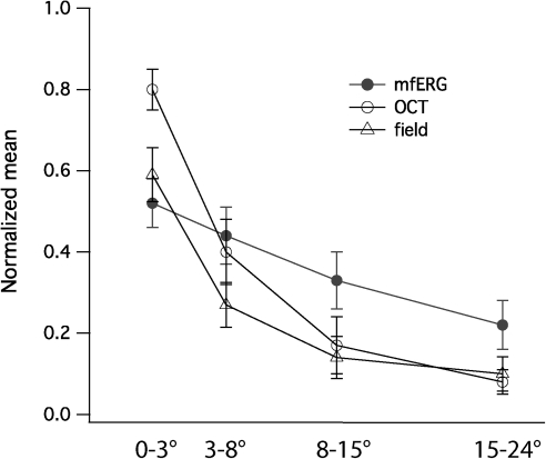
Figure 6.
Normalized mfERG amplitude, field sensitivity, and receptor layer thickness measure (ONL × OS) in the central 24°. Except in the central 3°, the three indices showed good correspondence in the characterization of photoreceptor degeneration in 10 patients with RP. In the central 3°, normalized mfERG amplitude showed significant larger reduction than SD-OCT thickness. Filled circle, mfERG amplitude; unfilled circle, SD-OCT measure; triangle, visual field sensitivity.

