Fig. 1.
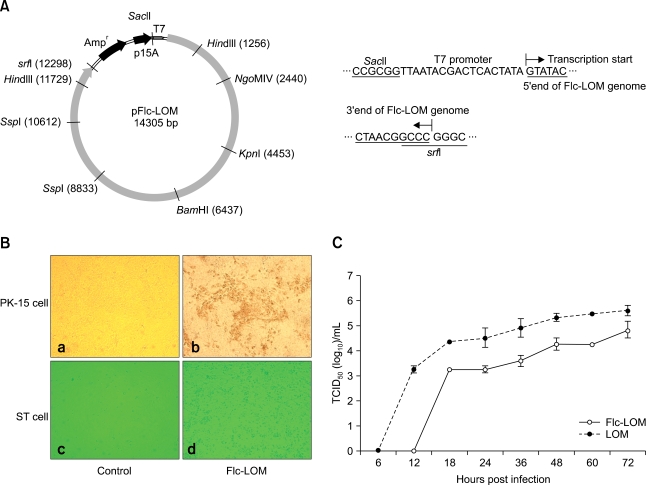
Strategies for construction and in vitro growth characterization of Flc-LOM. (A) Circular map of pFlc-LOM showing the relative positions of the LOM cDNA, ampicillin resistance gene, and P15A origin of replication. Locations of relevant restriction sites and the T7 promoter are indicated. The T7 RNA promoter was located immediately upstream of the 5' end of the genome, and a SrfI site was at the 3' end. The genome sequences are underlined. (B) Using the classical swine fever virus-specific monoclonal antibody LOM01, an indirect immunoperoxidase assay of Flc-LOM was performed in PK-15 cells 3 days after infection with a multiplicity of infection (MOI) of 1 (a and b, × 200). The Flc-LOM biotype was determined using the exaltation of Newcastle disease virus method. Serial dilutions from 101.0 to 106.0 of Flc-LOM were added to primary swine testicular cells. After 4 days, the supernatant was removed and the cells were infected with Newcastle disease virus (104.0 PFU/mL). A cytopathic effect was observed 3 days post-infection and photographed using an inverted microscope (c and d, × 200). (C) Replication kinetics of the Flc-LOM and LOM viruses. PK-15 cells seeded in T25 flasks were infected at a MOI of 1. The data are from three independent experiments. Virus titers are shown as log10 104 median TCID50/mL. ST: swine testicular.

