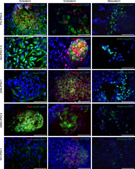
Figure 5.
EB-based differentiation of iPSC lines reveals three lineage-specific marker expressions. Disease-specific and wild-type iPSC lines were spontaneously differentiated in hESC medium without bFGF for the formation of EBs. Two weeks later, EBs were attached on a gelatin coated dish and allowed to differentiate into all three germ layers for 10 days. Immunostaining was performed using antibodies specific to ectoderm (Nestin, green; Pax6, red), endoderm [AFP (α-fetoprotein), green; HNF3β, red], and mesoderm (Brachyury, green) lineages. Scale bars = 100 µm.

