Fig. 1.
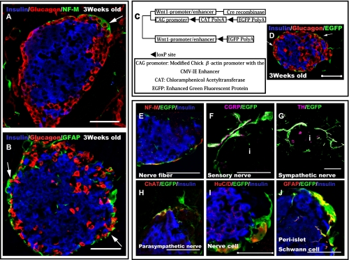
Neural crest (NC) derivatives surrounding islets differentiated into neural-related cells and tended to be distributed in closer proximity to alpha cells. (A and B) Three-week-old mouse cells stained with neurofilament (NF-M) (green) (A), glial fibrillary acidic protein (GFAP) (green) (B), insulin (blue) (A and B), and glucagon (red) (A and B) antibodies, showing immunoreactivity in regions surrounding the islets. Most of the nerve-related cells tended to be located in closer proximity to alpha cells than to beta cells (arrows). (C) Constitutive expression of enhanced green fluorescent protein (EGFP) in NC cells and their derivatives in double transgenic Wnt1-Cre/Floxed-EGFP mice. (D) Pancreatic region of a 3-week-old Wnt1-Cre/Floxed-EGFP mouse; the cells were stained with insulin (blue), glucagon (red), and GFP (green) antibodies. EGFP immunoreactivity is observed around the islets. Most NC derivatives tended to be located in closer proximity to alpha cells than to beta cells (arrows). (E–J) To determine the cell types of NC derivatives differentiated into around islets, immunostaining experiments were performed with various nerve-related and EGFP antibodies. Neural fibers (sympathetic nerves (G), parasympathetic nerves (H), and sensory nerves (F)) surrounding the islets were found to be derived from NC cells. Moreover, pancreatic ganglion cells (I) that made contact with islets were also found to be derived from NCs. peri-islet Schwann cells (PISCs) (J) surrounding the islets were also derived from NC cells. Bar=50 µm for all Figures. i=Islet.

