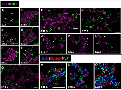
Fig. 2.
Distribution of NC derivatives during pancreatic development. (A–L) Immunohistochemical staining of pancreatic regions of Wnt1-Cre/Floxed-EGFP mouse embryos at each embryonic stage. Staining was performed with pancreatic and duodenal homeobox 1 (PDX1) antibody (magenta) and GFP antibody (green). (A) At embryonic day 9.5 (E9.5), Wnt1-Cre/Floxed-EGFP embryos did not have any NC derivatives in close proximity to the ventral and dorsal pancreatic buds (arrow). (B) At E10.5, NC derivatives were observed in a close proximity to both pancreatic buds. They were closer to the dorsal pancreatic bud than to the ventral pancreatic bud (arrowheads). (C and D) From E11.0, NC derivatives surrounded PDX1-positive cells. (E) At E12.5, as branching of buds progressed, NC derivatives were distributed along the pancreatic epithelial cells. (F) At E14.5, several NC derivatives tended to be located close to cells with high PDX1 expression (arrows). (J–L) Higher magnification image of the region in G–I, where NC derivatives were distributed in close proximity to PDX1-positive cells (arrows). (M–O) Immunohistochemical staining of pancreatic regions of mouse embryos is shown; staining was performed with insulin antibody (blue), glucagon antibody (red), and PDX1 antibody (green). During later stages of pancreatic development, PDX1-positive cells were mainly localized in insulin-positive rather than glucagon-positive cells. Bar=100 µm (B–O). VB=ventral pancreatic bud, DB=dorsal pancreatic bud.

