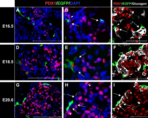
Fig. 3.
Glucagon-positive cells tended to be located between PDX1-positive cells and NC derivatives. (A–H) Immunohistochemical staining of pancreatic regions of Wnt1-Cre/Floxed-EGFP mouse embryos during later stages of pancreatic development. Staining was performed with PDX1 antibody (red) and GFP antibody (green), and nuclei were stained with DAPI solution (blue). (C, F, and I) Immunohistochemical staining results with PDX1 antibody (red), GFP antibody (green), and glucagon antibody (white). Figures B, E, and H are higher magnification images of A, D, and G, respectively. NC derivatives were distributed in closer proximity to PDX1-negative cells (arrowheads) than to PDX1-positive cells (arrows). Figures C, F, and I represent the same sections as B, E, and H, respectively, but were stained with glucagon antibody (white). PDX1-negative cells located in close proximity to NC derivative cells were glucagon-positive (arrows). Most NC derivatives tended to be distributed in closer proximity to glucagon-positive cells than to PDX1-positive cells (arrowheads) during later stages of pancreatic development. Bar=50 µm (A, D, G, C, F, and I).

