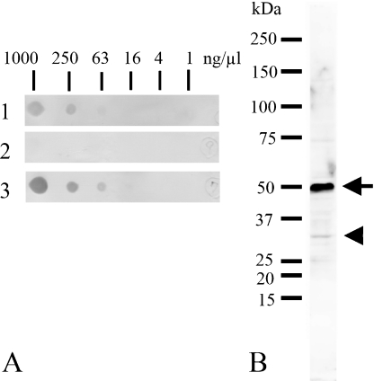
Fig. 4.
Immunospot test (A) of antigenic peptide detected by the antiserum (1, diluted at 1:10,000), unbounded fraction (2, diluted at 0.5 µg/ml) and purified antibody (3, diluted at 0.5 µg/ml), and Western blot analysis (B) of rat brain homogenates probed with the purified antibody (diluted at 0.5 µg/ml). A: Antibodies pre- (1) and post- (3) purification using affinity chromatography of antigenic peptide detect the peptide at a concentration of 25 ng/µl. Unbound fraction cannot detect the antigen (2). B: The purified antibody detected two bands with molecular weights of approximately 30 kDa (arrowhead) and 50 kDa (arrow).

