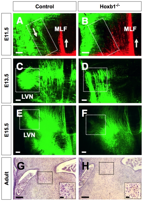
Figure 2. The projection pattern of the lateral vestibulospinal tract is missing Hoxb1−/− embryos.
(A, B) Fluorescent anterograde labeling of E11.5 control and Hoxb1−/− embryos injected with NeuroVue maroon (green) in the intermediate region of r4 and NeuroView (red) on the medial side of the caudal hindbrain to label the medial longitudinal fasciculus (MLF). Note prominent labeling of the axons emanating from the intermediate region of r4 is absent in the Hoxb1−/− embryo. (C, D) Fluorescent retrograde labeling of E13.5 control and Hoxb1−/− embryos injected with NeuroVue maroon (green) on the ipsilateral side of the spinal cord. Note prominent labeling of the LVN neurons in the control, but absent in the Hoxb1−/− embryo. (E, F) Fluorescent retrograde labeling of E15.5 control and Hoxb1−/− embryos injected with NeuroVue maroon (green) on the ipsilateral side of the spinal cord. Note prominent labeling of the LVN neurons in the control, but absent in the Hoxb1−/− embryo. (G, H) Transverse section through the lateral vestibular nucleus (LVN) in adult control and Hoxb1−/− hindbrain stained for hematoxylin and eosin. Lower right inset shows high magnification of the outlined area of the LVN. Large multipolar neurons are prominent in the LVN area of the control mouse, whereas only small to medium sized neurons are observed in the LVN area of the Hoxb1−/− mouse. Scale bars (A–F = 100 µm; G, H = 250 µm; G, H insets = 50 µm).

