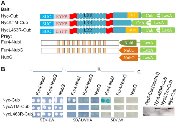
Figure 4. Genetic analysis shows murine nyctalopin has a single transmembrane domain.
A. Schematic diagram of bait and prey constructs used to dissect the topology of nyctalopin. In the schematic, the blue rectangle represents the invertase signal sequence (SUC), the grey rectangle EYFP, the aqua ovals each LRR domain, the aqua rectangle with predicted transmembrane (TM) domain (orange rectangle) the C-terminus of nyctalopin, the chevron the C-terminus of ubiquitin (Cub) and the green rectangle the artificial transcription factor VP16LexA. NycΔTM-Cub has amino acids 455–476 (containing TM1) deleted from nyctalopin. NycL463R-Cub has a base substitution that creates a leucine to arginine substitution at position 463 of nyctalopin. Bi. Shows that all bait and prey plasmids were present in the NMY32 yeast strain and supported growth on the selective SD/-LW plates. Bii. The growth tests for interaction of bait and prey fusion proteins when the transformants are grown on SD/-LWHA plates. Biii. An X-gal assay for expression of β-galactosidase confirming the interaction in Bii. These results support the conclusion that only the Nyc-Cub/Fur4-NubI combination interact, indicating that there is a transmembrane domain in nyctalopin and that the C-terminus of nyctalopin is cytoplasmic. C. Western blot showing truncated transmembrane and L463R mutants are expressed in NMY32 yeast strain.

