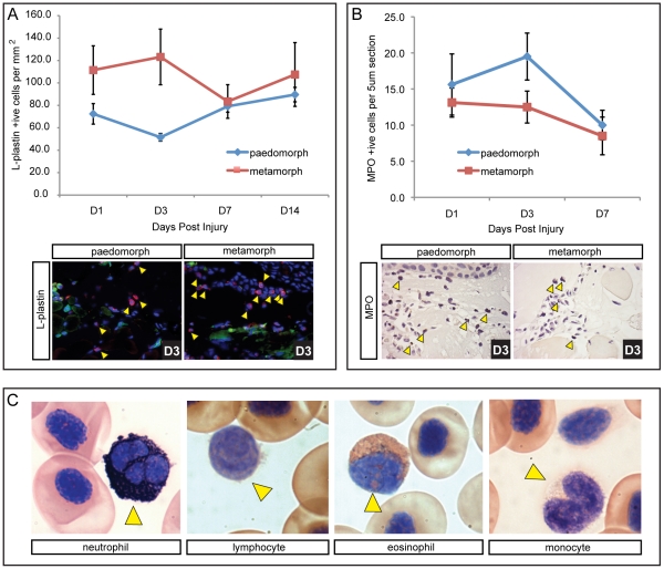
Figure 7. Higher initial leukocyte levels are present in terrestrial axolotls coincident with slower re-epithelialization, but neutrophil levels are not different between morphs.
L-plastin was used as a pan-leukocytic marker to quantify total leukocyte levels following injury. A) Total leukocytes counted per mm2 in the wound bed at 1, 3, 7 and 14 dpi (n = 4 for each morph). L-plastin positive cells are red (yellow arrows), nuclei are stained blue (Hoescht) and green fluorescence was used to account for autofluorescencing erythrocytes that were excluded as leukocytes. An influx of leukocytes was observed 24 hrs dpi, with higher numbers present in terrestrial axolotls. Leukocyte levels dropped in metamorphs at D7 and converged with paedomorph levels. Levels for paedomorphs were not significantly changed after D1. B) All neutrophils present in the wound bed (yellow arrows) were counted on 5 µm sections above the muscle using myloperoxidase (n = 8 for each morph; see Figure S6 for positive control staining in liver). Neutrophil levels were generally low and were not significantly different between paedomorphs and metamorphs, although they did drop significantly at D7. C) Modified Wright-Giemsa stain used to indentify individual leukocytes in circulating axolotl blood. Sudan black was used to stain neutrophils. Yellow arrows indicate the specific leukocyte type.

