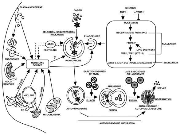
Figure 1.
Schematic model of dynamic membrane events contributing to autophagosome life and death. The life and death of autophagosomes, which is evolutionarily conserved, involves the following stages (underlined): initiation, phagophore nucleation, phagophore elongation through two conjugation cascades, cargo selection, sequestration and packaging, membrane recycling regulated by Atg9, fusion with lysosomes, degradation and efflux. Key proteins that act at these stages during such processes are listed. Potential sources for the phagophore include the endoplasmic reticulum (ER), nuclear membranes, mitochondria, plasma membrane, Golgi complex and endosomes. The phagophore expands, sequestering and packaging the selected autophagy cargo, and finally closes, forming an immature autophagosome. During the maturation process, an amphisome is formed from the fusion of an autophagosome with early endosomes or multivesicular bodies, whereas an autolysosome/autophagolysosome is formed as a product of fusion of amphisomes events with late endosomes or lysosomes. Once in the lysosomes, the autophagic cargo is degraded into essential building blocks that are transported back into the cytoplasm. Abbreviations: AMPK, AMP-activated protein kinase; Atg, autophagy-related; BECLIN 1, BCL-2 interacting myosin/moesin-like coiled-coil protein 1; ER, endoplasmic reticulum; ER/ENDO, endoplasmic reticulum/endosomes contact; ER/MITO, endoplasmic reticulum/mitochondria contact; LC3, light chain 3; mTORC1, mammalian target of rapamycin complex 1; MVBs, multivesicular bodies; PM, plasma membrane; PtdIns3P, phosphatidylinositol 3-phosphate; PtdIns3KC3; phosphatidylinositol 3-kinase class III; ULK1, UNC51-like kinase 1; WIPI1/WIPI2, WD repeat domain phosphoinositide-interacting 1/2.

