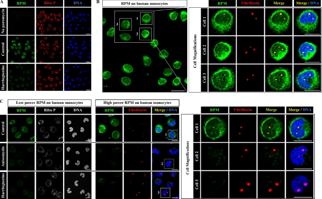
Figure 6.
Nuclear RPM staining in monocytes. (A) Elutriated human peripheral blood monocytes pretreated or not pretreated with harringtonine 15 min before RPM staining (5-min PMY pulse in the presence of emetine) were fixed and extracted simultaneously with polysome buffer containing PFA and digitonin. Note that in the absence of PMY no RPM staining is visible. (B) Same PMY staining protocol as in A. Intense RPM staining clearly colocalizes with nucleoli (red), which are smaller in monocytes than HeLa cells as shown by fibrillarin staining. (C) Elutriated monocytes pretreated or not pretreated with harringtonine or anisomycin for 15 min before RPM staining were fixed and extracted simultaneously with polysome buffer containing PFA and digitonin. RPM staining in cytoplasm and nucleoli is blocked by either inhibitor as seen in increasing magnifications from left to right, demonstrating the dependence of ribosome-catalyzed puromycylation of the nascent chain. Bars: (A and B [main image]) 20 µm; (B [magnification] and C) 10 µm. Ribo P, ribosomal P.

