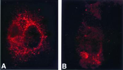
Figure 2.
Intracellular distribution of IL-15 in CV-1 cells infected with recombinant vaccinia viruses. CV-1 cells were infected with the respective recombinant viruses, and the expression profile of IL-15 was examined by confocal microscopy 18 h after infection. In A, cells were infected with IL-15(NS) vaccinia virus, and in B, cells were infected with IL-15(IS) vaccinia virus.

