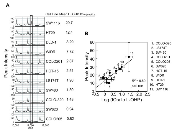
Figure 1.
L-OHP sensitivity and candidate peak selection. (A) Protein expression profiles of each cell line on CM10 array at pH 4.5. The candidate peak is enclosed by the rectangle. (B) Peak intensity of the 11.1 kDa protein in 11 CRC cell lines strongly correlates with L-OHP sensitivity. The peak intensity and IC50 value of each cell line are plotted as means ± S.D. (peak intensity, n = 3; IC50, n = 3 or 4).

