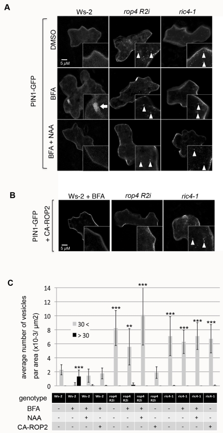
Figure 3. The ROP2-RIC4 pathway mediates auxin-induced inhibition of PIN1 endocytosis.
PIN1-GFP was transiently expressed for 24 h alone (A) or coexpressed with CA-ROP2 construct (B) in Arabidopsis PCs of WT control (Ws-2), rop4 R2i, or ric4-1 leaves treated with DMSO (mock control), BFA, or BFA plus NAA. Medial sections of confocal images were taken, and internalized PIN1-GFP structures (BFA body-like, larger than 30 pixels; Ara7 vesicle-like, smaller than 30 pixels) were analyzed. (A) PIN1-GFP vesicles (Ara7 vesicle-like, see Figure S4) were rarely found in untreated Ws-2 cells and were abundant in rop4 R2i or ric4-1 cells. BFA (50 µM) treatment induced large aggregation of PIN1-GFP structures into BFA bodies in Ws-2 cells, but did not affect PIN1-GFP vesicles in rop4 R2i or ric4-1 cells. Application of 100 nM NAA (BFA+NAA) inhibited the accumulation of PIN1-GFP in BFA bodies in Ws-2, but did not alter the formation of PIN1-GFP vesicles in rop4 R2i or ric4-1 cells. (B) CA-ROP2 expression prevented PIN1-GFP accumulation in BFA bodies in Ws-2. CA-ROP2 also suppressed the formation of PIN1-GFP vesicles in rop4 R2i but not in ric4-1. (C) Quantitative analysis of PIN1-GFP containing BFA bodies and vesicles shown in (A) and (B). Vesicles were categorized by pixel area in the images into two classes (see Figure S5): <30 = smaller than 30 pixels, similar to the size of Ara7 vesicles; >30 = larger than 30 pixel, BFA bodies. Error bars represent SD (n = 10). p-Values (against Ws-2–untreated cells) were determined by two-tailed Student's t test assuming equal variances (**, p<0.01; ***, p<0.001).

