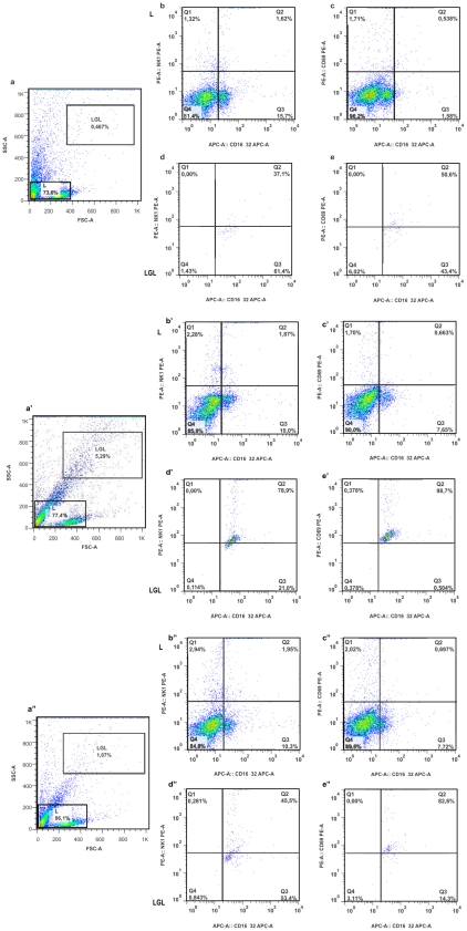
Figure 6. Monitoring presence of activated NK cells in bloodstream post amastigote intraperitoneal inoculation.
Flow cytometry was performed with mononuclear cells prepared from mice left without any inoculation (a, b, c, d and e), and mice that were given intraperitoneal G strain amastiogotes at day-8 post-inoculation (a′, b′, c′, d′ and e′) at day-25 (b″, c″, d″ and e″). Note the higher percentage of activated NK cells at day-8 post-inoculation (p<0.01). Gates: L – lymphocytes and LGL – large granular lymphocytes (NK cells).

