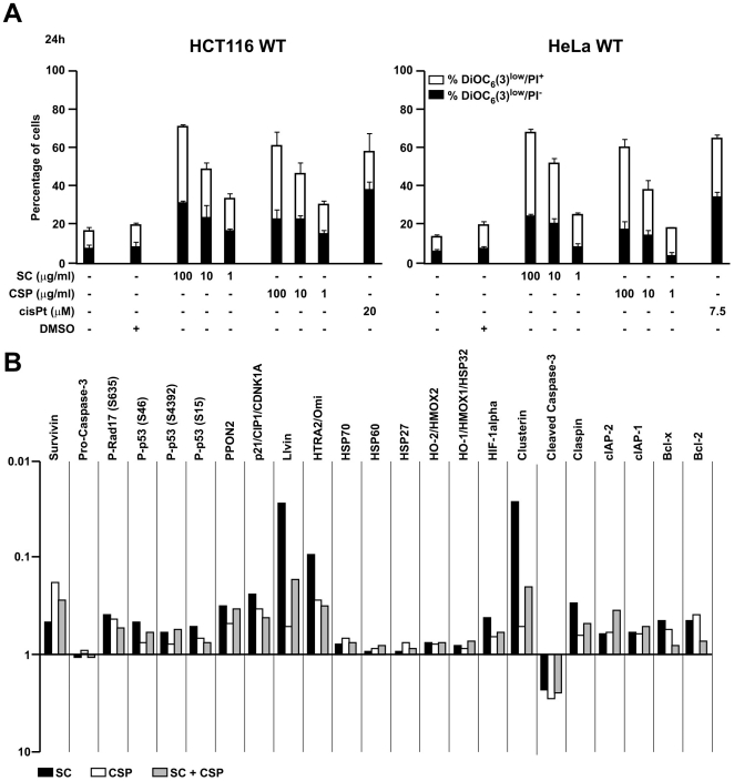
Figure 4. Effect of SC and CSP on apoptosis induction.
Detection of dead and dying cells by FACS (A). Cells treated overnight with SC, CSP and cisPt were stained with ΔΨm-sensitive dye DiOC6(3) and the vital dye propidium iodide (PI). The white portions of the columns refer to the DiOC6(3)low/PI+ population (dead) and the remaining part of the column corresponds to the DiOC6(3)low/PI- (dying) population. Results are means ± S.E.M. of three independent experiments. Expression profile of apoptosis-related proteins (B). Cell lysates from HeLa cells, treated with SC (10 µM) and CSP (10 µM) overnight, were analyzed by human apoptosis profiler. Data are expressed as means ± S.D. of two independent experiments.

