Fig. 4.
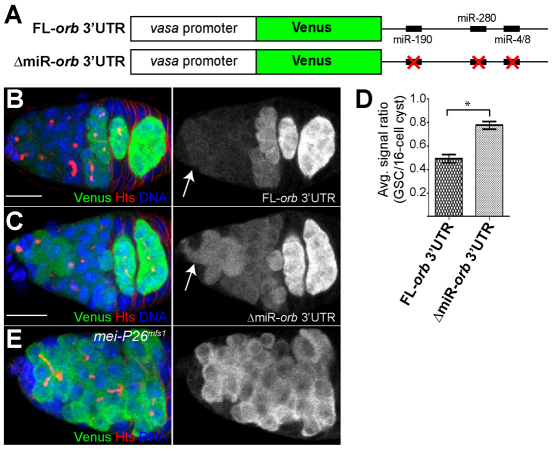
The 3′UTR of orb mRNA contains miRNA binding sites and responds to changes in Mei-P26 levels. (A) The reporter genes used to evaluate the importance of predicted miRNA binding sites within the 3′UTR of orb mRNA. (B,C) Full-length orb 3′UTR (FL-orb3′UTR) (B) and an orb 3′UTR reporter that contains mutated miRNA binding sites (ΔmiR-orb3′UTR) (C) stained for Venus (green), Hts (red) and DNA (blue). The FL-orb3′UTR reporter exhibits very low levels of expression in GSCs and high levels in 16-cell cysts, similar to the Hsp83-lacZ-orb3′UTR reporter and Orb protein (see Fig. 3C). By contrast, the ΔmiR-orb3′UTR reporter displays elevated levels of expression in GSCs. The arrows point to GSCs. (D) The average signal ratio between Venus expression in GSCs and that in 16-cell cysts for the indicated reporters (n=11; germaria from at least two different transgenic lines were evaluated for each reporter; *P<0.005). Error bars indicate s.d. (E) Cells adjacent to the cap cells express the FL-orb 3′UTR reporter in a mei-P26 mutant background. Scale bars: 10 μm.

