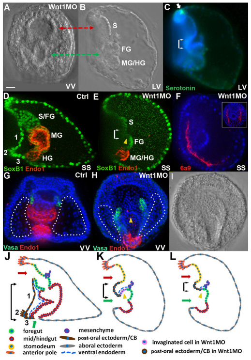
Fig. 1.
The structure of sea urchin larvae lacking Wnt1. The Wnt1 morphant phenotype was produced consistently with either of two morpholinos (see Materials and methods). The dose used for each morpholino was chosen to yield a high rate of embryo survival and a consistent phenotype that was observed in >95% of embryos in at least 20 batches of embryos. (A,B) A Wnt1 morphant at the 4-day larval stage, ventral (A) and lateral (B) views, showing a large indentation in the stomodeal region (red arrow) and the blastoporal opening (green arrow) in the same ventral plane. (C) Lateral view of the embryo in B, stained for serotonin (green, white arrow) and nuclei (DAPI, blue). (D,E) Control and Wnt1 morphant 4-day larvae stained for SoxB1, which is present in nuclei of ectoderm and foregut (green), and Endo1 (red), which marks mid- and hindgut. (F) A 3-day Wnt1 morphant in which the 6a9 monoclonal antibody (red) labels primary mesenchyme cells that secrete the skeletal spicules; DAPI (blue), lateral view. Inset: oral view showing both spicules. (G,H) Control larva (G) and Wnt1 morphant (H), ventral views, stained for Vasa (green), which labels the small micromere descendants that populate the coelomic pouches of larvae, Endo1 (red) and DAPI (blue). Dotted line indicates posterior-ventral ectoderm. (I) DIC image of the larva in H, showing that the blastoporal opening is cup-shaped and joins a trough of endodermal tissue that is continuous with the oral ectoderm and is open to the medium. (J-L) Schematic of tissues in control embryos (J) and Wnt1 morphants (K,L). Double arrows in C,E,J-L indicate the posterior ventral corner (PVC) region. Yellow arrowheads in E,H,K,L mark invaginated tissue from the ventral side of the embryo. Green arrows indicate the blastopore. Red arrows indicate the stomodeal region. CB, ciliary band; FG, foregut; HG, hindgut; LV, lateral view; MG, midgut; S, stomodeum; SS, sagittal section; VV, ventral view; Wnt1MO, Wnt1 morpholino; 1, post-oral ectoderm; 2, post-oral transverse CB; 3, supra-anal ectoderm. Scale bar: 20 μm.

