Fig. 6.
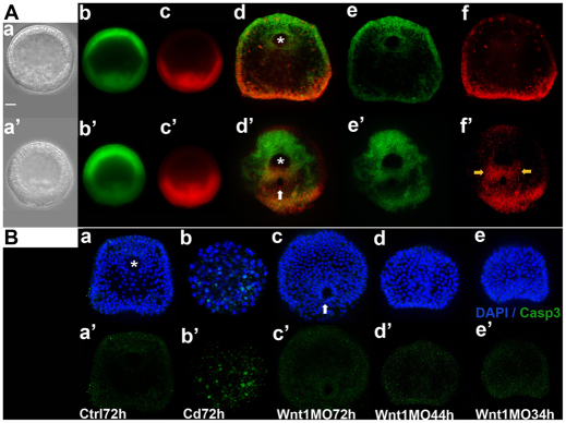
The altered PVC does not result from extensive cell loss in Wnt1 morphants. (A) PVC progeny are present in the ectoderm between the blastoporal and stomodeal regions in 3-day old Wnt1 morphants. The posterior half of the embryo was labeled red by photoactivating KikGR at early mesenchyme blastula stage (control embryo, Aa-c; Wnt1 morphant, Aa′-c′) and photographed at 3 days post-fertilization to monitor the positions of red cells in the PVC (same control embryo, Ad-f; same Wnt1 morphant, Ad′-f′). The yellow arrows in Af′ indicate the PVC progeny between the blastopore and stomodeal regions. (B) No caspase 3 (green) is detected in Wnt1 morphants. (Ba,a′) Control 3-day embryo; (Bb,b′) cadmium chloride-treated 3-day embryo; (Bc-e,c′-e′) Wnt1 morphants at the indicated times. Asterisks mark mouth in (Ad,Ba) and stomodeal ectoderm in (Ad′). White arrows indicate blastopore in (Ad′,Bc). Scale bar: 20 μm.

