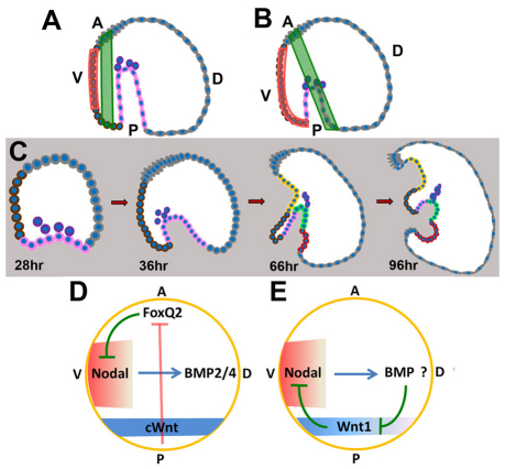
Fig. 7.
Model: Wnt1-mediated suppression of nodal expression maintains patterning orientation along the DV axis. (A,B) Diagrams of the expansion of oral ectoderm (orange) and the shift of the CB (green) with respect to the AP axis in control (A) and Wnt1 morphant (B) embryos. In B, Nodal expression (orange) extends ectopically toward the blastopore. (C) Time course of development from mesenchyme blastula (28-hour), gastrula (36-hour), early pluteus larva (66-hour) and late pluteus larva (96-hour) stages in Wnt1 morphants, illustrating positions of different cell types in a mid-sagittal section. Color codes are given in Fig. 1. (D) Initiation of AP and DV axial patterning by Wnt/β-catenin and Nodal signals and interactions between these signals at early stages. Wnt/β-catenin eliminates FoxQ2 from all but the anterior-most ectoderm. FoxQ2 suppresses expression of nodal, which, along with its target gene, bmp2/4, is necessary and sufficient for DV axial patterning. This double-negative regulatory device coordinates early AP and DV axis patterning. (E) Interactions between Wnt1 and Nodal signaling during gastrulation. Nodal-dependent processes, possibly working through BMP2/4, eliminate Wnt1 expression from dorsal posterior cells but not from those on the ventral side. Conversely, ventral Wnt1-dependent processes suppress nodal expression in posterior ventral cells, which is required to maintain the correct orientation of patterning along the DV axis. A, anterior; D, dorsal; P, posterior; V, ventral.

