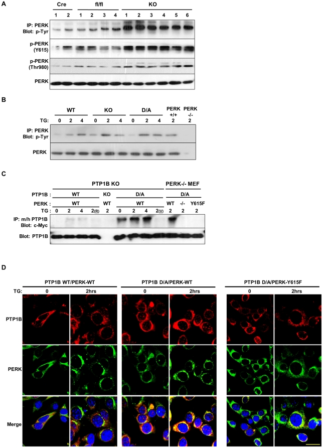
Figure 3. PTP1B dephosphorylation of PERK.
(A) PERK was immunoprecipitated from BAT lysates of Cre, fl/fl and adipose-PTP1B KO (KO) male mice fed HFD for 30 weeks, and then immunoblotted with anti-phosphotyrosine antibodies, phospho-specific antibodies for PKR Tyr293 (PERK Y615) and PERK (Thr980). Blots were also probed for PERK to control for loading. Each lane represents brown adipose tissue from a different animal. (B) Differentiated WT, KO and D/A brown adipocytes and PERK−/− and PERK+/+ fibroblasts were treated with thapsigargin (TG) for the indicated times. Immunoprecipitates of PERK were immunoblotted with anti-phosphotyrosine antibodies. Blots were also probed for PERK to control for loading. (C) PTP1B KO preadipose cells were co-transfected with PTP1B WT and substrate-trapping D181A mutant (D/A) and Myc-tagged PERK wild type. In addition, PERK KO fibroblasts were co-transfected with PTP1B D/A and Myc-tagged PERK WT and Y615F mutant. Cell were treated with TG for the indicated times then lysed in NP40 or RIPA (R), with or without pervanadate (V) treatment. Lysates were immunoprecipitated using mouse (m) and human (h) PTP1B antibodies and immunoblotted using anti-c-Myc and anti-(m/h) PTP1B antibodies. (D) PTP1B KO brown preadipocytes were transfected with hPTP1B WT, or hPTP1B D/A (red) and c-Myc-PERK WT or Y615F mutant (green), treated with thapsigargin (TG) for 2 hours and visualized by fluorescence confocal microscopy. Scale bar corresponds to 20 µm.

