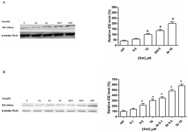
Figure 1. Somatostatin induces an increase of IDE expression in microglia cells.
Western blot analysis of normalized lysis samples from rat primary microglia (A) and BV-2 (B) indicates that IDE level increases after 24 hrs of somatostatin incubation, while the internal control ß-tubulin is constant. The additional incubation with sst after 6 hrs from first round strengthens the effect on IDE expression (left panel). Densitometric analysis of IDE WB signals, average ± ES of 5 independent experiments in triplicate (right panel). *P<0.05, one-way ANOVA, followed by Tukey's test, n = 15.

