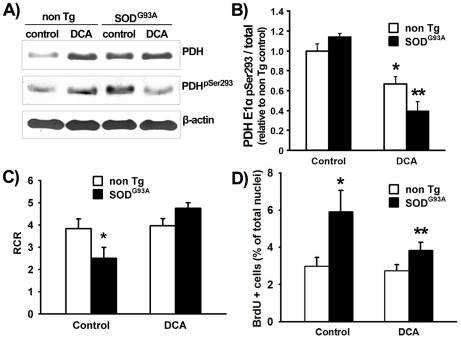
Figure 2. DCA recovers mitochondrial respiration rate and controls proliferation in SOD1G93A astrocytes.
A) Representative immunoblot for PDH-E1α(pSer293), total PDH-E1α, and β-actin of lysates from non Tg and SOD1G93A astrocytes after 24 h treatment with DCA or vehicle as described in Methods. B) Quantification of the PDH-E1α(pSer293) to total PDH-E1α ratio between relative densitometric levels normalized against vehicle-treated non Tg astrocytes. C) Calculated respiratory control ratio (RCR) for mitochondria from non Tg or SOD1G93A-bearing astrocytes treated with DCA or vehicle as indicated. D) Percentage of BrdU immunoreactive nuclei of non Tg and SOD1G93A astrocytes after 24 h treatment with DCA. Data for panels B, C, and D are expressed as mean ± SEM from three independent experiments performed in duplicate. *p<0.05, significantly different from non Tg control. **p<0.05, significantly different from SOD1G93A control.

