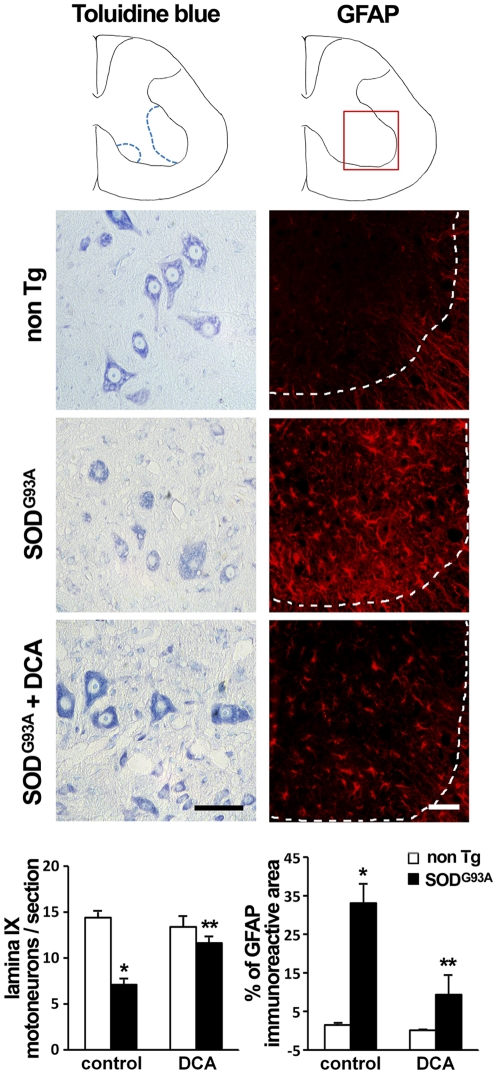
Figure 7. DCA reduces motor neuron loss and astrocyte reactivity in the spinal cord of SOD1G93A mice.
Representative Toluidine blue stain (left column) and GFAP immunofluorescence (red, right column) in anterior horn spinal cord sections from non Tg (top), SOD1G93A control (middle) or DCA-treated SOD1G93A (bottom) mice. Dotted lines in right column panels indicate the limit between grey and white matter. The graphs indicate the number of neuronal somas located in Rexed lamina IX (left) and the percentage of GFAP immunoreactive area in the ventral horn (right) in the indicated groups of animals. The corresponding measurement areas are drawn in the top. Data are mean ± SEM from at least three animals per group as indicated in Methods. *p<0.05, significantly different from non Tg control, **p<0.05, significantly different from SOD1G93A control. Scale bars: 50 µm.

