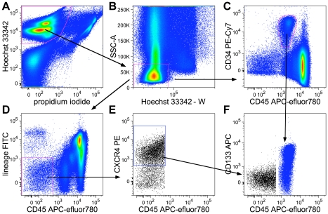
Figure 1. Gating of VSEL cells and CD34+CD45dim cells in hUCB.
hUCB TNC were isolated by NH4Cl lysis and stained with fluorescent antibodies, Hoechst 33342, and PI. A) Gating of living (PI−) nucleated (Hoechst 33342+) cells; B) pulse processing: exclusion of doublets (Hoechst-Whigh) and granulocytes (SSC-Ahigh). C) Gating of CD34+CD45dim Hoechst 33342+PI−. D) Gating of CD45−Lin− cells. E) Gating of CD45−Lin− CXCR4+Hoechst 33342+PI− VSEL cells. F) Expression of CD133 in CD34+CD45dim cells (blue) and VSEL cells (black).

