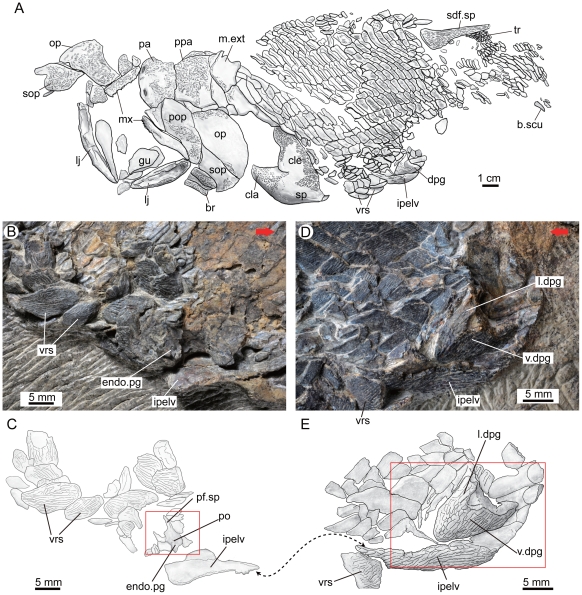
Figure 2. The holotype (V15541) of Guiyu oneiros Zhu et al., 2009.
A. Interpretative drawing of the part to show the position of the newly identified left pelvic girdle with dermal and endoskeletal components. B–C. Close-up of the counterpart to show the endoskeletal pelvic girdle in internal view (B) and interpretative drawing (C). D–E. Close-up of the part to show the dermal pelvic girdle in lateral view (D) and interpretative drawing (E). Red arrows point to the anterior end of the fish. The red rectangles indicate the close-up areas in Figure 3A and Figure 3B. The double arrows point to the corresponding positions of the fractured interpelvic plate in part (E) and counterpart (C). Abbreviations: br, branchiostegal ray; b.scu, basal scute; cla, clavicle; cle, cleithrum; dpg, dermal pelvic girdle; endo.pg, endoskeletal pelvic girdle; gu, gular; ipelv, interpelvic plate; l.dpg, lateral lamina of dermal pelvic girdle; lj, lower jaw; m.ext, median extrascapular; mx, maxillary; op, opercular; pa, parietal shield; pf.sp, pelvic fin spine; po, foramina for pterygial nerves and vessels; pop, preopercular; ppa, postparietal shield; sdf.sp, second dorsal fin spine; sop, subopercular; sp, pectoral fin spine; tr, lepidotrichia; v.dpg, ventral lamina of dermal pelvic girdle; vrs, ventral ridge scale.

