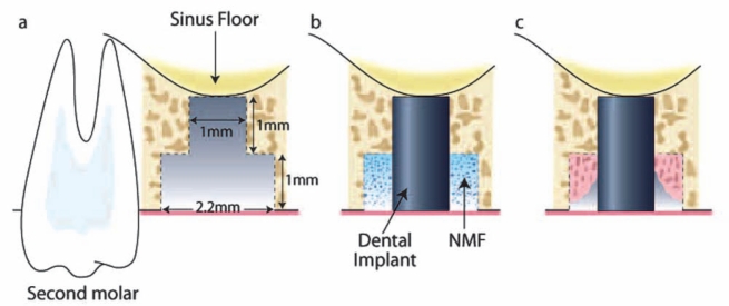
Figure 2.
Infrabony peri-implant defect in the rat. (a) After extraction of the maxillary first molar, the socket was allowed heal for ~ 4 wks. (b) During the second surgery, a bone defect was created by means of an osteotomy in the location of the former tooth. An implant (1 mm x 2 mm) was press-fit into position, and the NMF was delivered to the peri-implant bone defect, followed by soft-tissue wound closure. (c) At multiple time-points (10, 14, and 21 days), tissue samples were harvested, and bone healing was evaluated.

