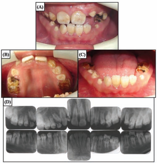
Figure 3.
Clinical photographs and radiographs of the affected brother of the proband (IV:3) at age 12 yrs. (A) Frontal view. Overjet and overbite are minimal, showing the edge-to-edge bite. Anterior teeth have dark brown pigmented areas. (B) Maxillary occlusal view. Enamel breakdown can be seen in the permanent first molars and deciduous teeth. Newly erupting premolars do not have pigmentation. (C) Mandibular occlusal view. Anterior teeth have dark staining. (D) Full-mouth intra-oral radiographs. Enamel has reduced radiopacity, but normal thickness can be seen in the newly erupted premolars.

