
Figure 3.
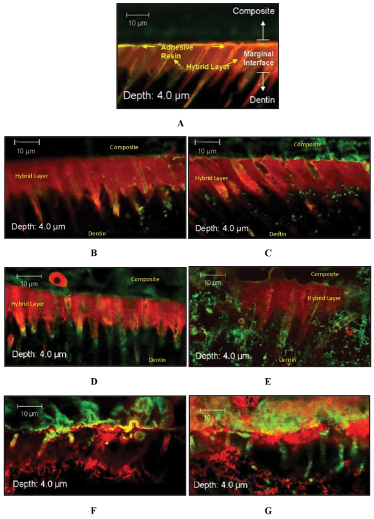
Selected Z-stack image series captured from interfacial margins of resin-dentin specimens assigned to either (A) non-incubated, (B) 7-day PBS incubation, (C) 7-day PCE+CE incubation, (D) 30-day PBS incubation, (E) 30-day PCE+CE incubation, (F) 90-day PBS incubation, or (G) 90-day PCE+CE incubation. Interfacial zones (composite, adhesive, hybrid layer, and dentin) are distinguishable in A and, to a lesser extent, in B-E; however, in F and G, the organization of these marginal components is disrupted. Resin impregnation of dentinal tubules in the hybrid layer is disrupted (F and G). Specimens were stained by means of a Live/Dead Baclight Viability Kit (magnification X62, 2X zoom). Live cells indicated by green fluorescence through interaction with Syto9; dead cells indicated by red fluorescence through interaction with propidium iodide.