
Figure 4.
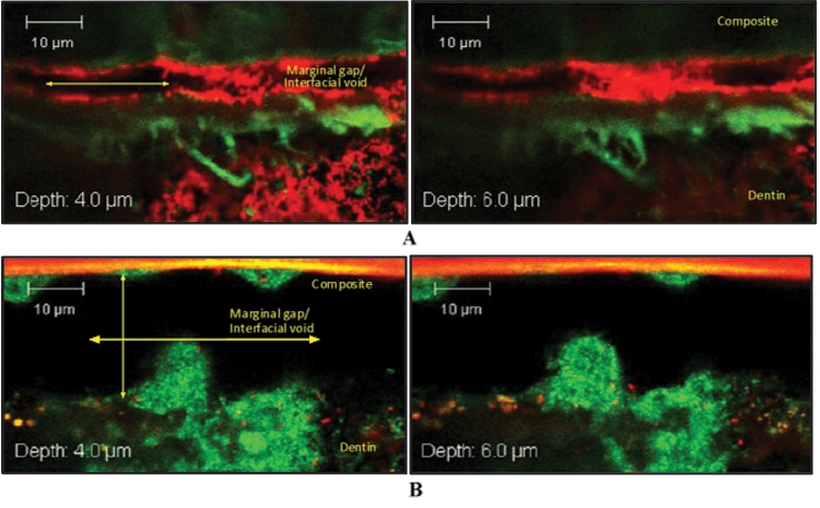
Selected Z-stack image series captured at interfacial ROIs of 2 90-day PCE+CE-incubated resin-dentin specimens. (A) Interfacial void spanning approximately 4-5 µm in height. (B) Interfacial void spanning over 20 µm in height. Characteristic of three-dimensional biofilm growth are interstitial voids that can be seen among fluorescently stained S. mutans microcolonies. In (B), large mushroom-shaped biofilm structures are found colonizing both the top and bottom axial walls. Specimens were stained by means of a Live/Dead Baclight Viability Kit (magnification X62, 2X zoom). Live cells indicated by green fluorescence through interaction with Syto9; dead cells indicated by red fluorescence through interaction with propidium iodide.