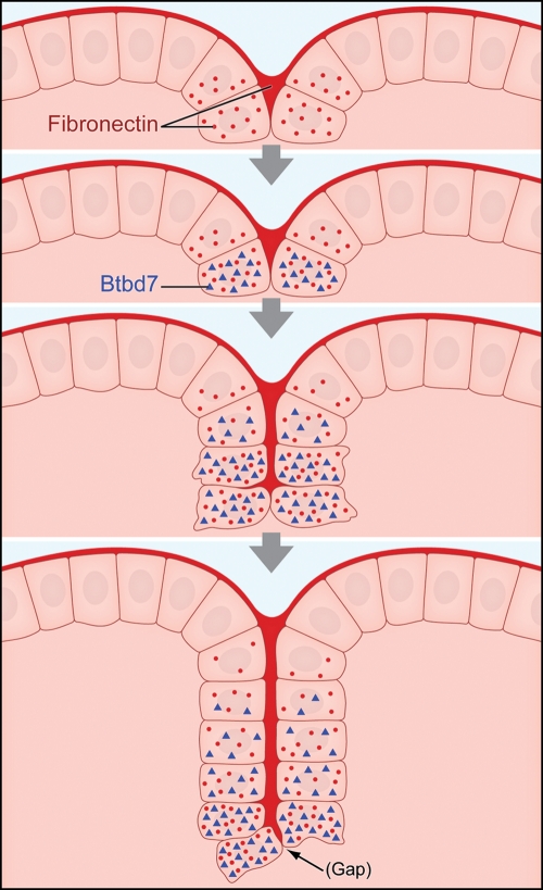
Figure 5.
Schematic representation of a new pathway regulating salivary gland cleft formation. The 4 images depict a cleft that initiates and then progresses by cleft deepening. The basement membrane at the basal surface of the peripheral epithelial cells and forming clefts both contain polymerized fibronectin (dark red), and salivary epithelial cells adjacent to the cleft synthesize new fibronectin (red dots). Fibronectin induces Btbd7. As the cleft progresses, Btbd7 decreases in cells near older (upper) parts of the cleft, as Btbd7 is mainly induced at the base of forming clefts. The cleft advances through progressive extension of the zone of fibronectin filling the cleft (Larsen et al., 2006) associated with the relatively stochastic formation of tiny gaps between cells expressing Btbd7. Average times between the second step (definitive cleft initiation) and the third step (advancing cleft) are approximately 2-4 hours, and approximately 3-5 hours between the third and the fourth steps depicted (deep cleft).

