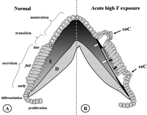
Figure 2.
Normal amelogenesis (A) and amelogenesis 24 hrs after an acute high exposure to fluoride (B). (A) Schematic drawing of normal amelogenesis in an imaginary cusp of a molar. D, dentin; E, enamel. Increasing greyness in enamel represents increasing mineral content. Successive stages of development from bottom to top. Aprismatic enamel with small crystallites is produced by early- and late-secretory ameloblasts. The bulk of (inner) enamel consists of prisms containing large crystals and is deposited by fully differentiated ameloblasts. (B) Amelogenesis 24 hrs after injection of 9 mg F/kg. Fluoride induces an (inner) hypermineralized layer (black line, white arrows) in fully secretory-stage enamel, running almost parallel to the surface of the enamel. It represents the mineralization front 24 hrs earlier at the time of fluoride injection. A later-formed (outer) layer is a hypomineralized layer (white line, black arrows); together, these lines form the double-response typical of fluoride. Two areas of intense hypermineralization are formed at both ends of these lines: one where the lines intersect the enamel-dentin junction in the inner aprismatic enamel, the other where the lines intersect the enamel surface with outer aprismatic enamel below late-secretory and transitional ameloblasts. Cyst formation occurs only under some groups of transitional ameloblasts (coronal cyst, coC) and early-secretory ameloblasts (cervical cyst, ceC) that detach from enamel surface. Maturation ameloblasts seem structurally unaltered. Fully secretory-stage ameloblasts recover completely after 24 hrs; only the double-response lines are reminiscent of the insult.

