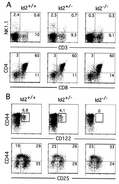
Figure 1.
Selective developmental defect in NK cells of the Id2−/− fetal thymus. (A) Cells of 17-dpc FT were stained with CD3-, NK1.1-, CD4-, and CD8-specific mAbs. Total thymocyte numbers were 1.5 ± 0.9 × 10⁶, 1.8 ± 0.6 × 10⁶ and 0.7 ± 0.6 × 10⁶ for Id2+/+, Id2+/−, and Id2−/− FT, respectively. (B) Cells of 14-dpc FT were stained with FITC-conjugated mAbs specific for TER119, Mac-1, Gr-1, B220, NK1.1, CD3, CD4, and CD8. Only FL-1-negative cells were analyzed for expression of CD44 vs. CD122 and CD44 vs. CD25. Total thymocyte numbers were 5.6 ± 1.4 × 10⁴, 3.2 ± 1.4 × 10⁴, and 1.5 ± 1.3 × 10⁴ for Id2+/+, Id2+/−, and Id2−/− FT, respectively. Numbers represent the percentage of cells in the gated population and in each quadrant in A and B.

