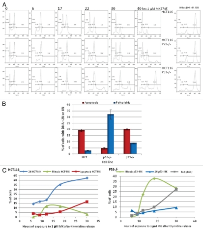
Figure 2.
Cell cycle effect and induction of apoptosis by MK8745 (5 µM) in isogenic variants of HCT-116 cells (parental, p53−/−, p21−/−). (A) Flow cytometry analysis of the HCT 116 (top) and its isogenic variants; p21−/− (middle) and p53−/− (bottom) upon exposure to MK for different time points (6, 17, 22, 30 and 40 h) and exposure to ABI (100 nM AZD 1152) for 40 h after Propidium Iodide staining. (B) HCT116, p53−/− and p21−/− cells treated with 5 µM MK for 24 h and analyzed for its DNA content after staining with Propidium Iodide by flow cytometry analysis. Polyploidy (8N, blue bars) and apoptosis (<2N, red bars). (C) Mitotic population determined by flow cytometry analysis after probing for phospho MPM2, a mitotic marker, in all the three cell lines. HCT116 (i) and p53−/− (ii) cells were double thymidine blocked and released to 1 µM MK, harvested at 6, 9, 12, 18 and 30 h and analyzed for its DNA content after staining with Propidium Iodide and mitotic population after phospho MPM2 staining by flow cytometry analysis. All the results are representative of 3–4 independent experiments.

