
Figure 2.
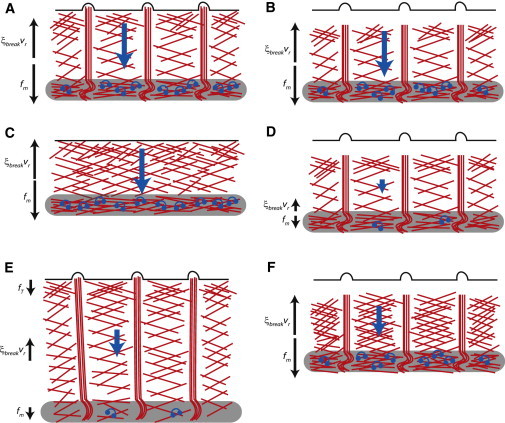
Schematics of growth-cone experiments used to constrain and test the model. (A) Under control conditions, myosin pulling is balanced by the actin breaking force. (B) When the leading-edge polymerization is inhibited by cytochalasin B, the force balance does not change. (C) When filopodia are removed with a low concentration of cytochalasin B, the force balance and flow do not change but the P domain shortens. (D) Inhibition of both polymerization (by cytochalasin B) and myosin (by blebbistatin) lowers both pulling and resistive forces and actin flow. (E) When myosin is inhibited by blebbistatin, the P domain widens and the remaining actin flow is driven by a combination of leading-edge tension and weakened myosin contractile force. (F) Inhibition of actin depolymerization by jasplakinolide increases the resistance of the actin network and slows the actin flow.