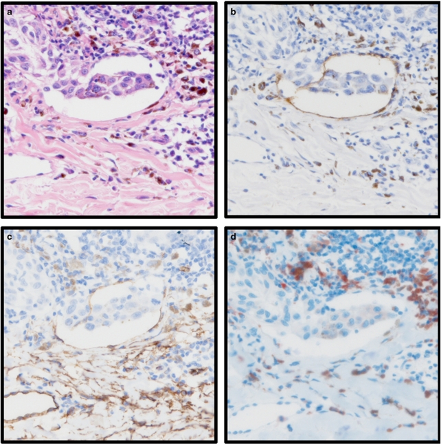
Figure 1.
Photomicrographs of tissue stained with D2-40 (lymphatic vessels), anti-CD34 (pan-vascular marker), anti-CD68 (macrophage marker), and haematoxylin and eosin (H&E) from two tumours (a–d tumour 1, c–h tumour 2). Examples are given of a true positive determined in H&E-stained tissue (tumour 1) and an example of a false negative in H&E-stained tissue (tumour 2). Tumour one; (a) vessel invasion determined in H&E-stained tissue; (b) invasion occurs in D2-40-stained lymphatic vessel; (c) invasion is visible in a vessel stained with anti-CD34; (d) anti-CD68-stained macrophage are observed around the invasion. Tumour 2; (e) vessel invasion not visible in H&E-stained tissue; (f) invasion occurs in D2-40-stained lymphatic vessel; (g) consecutive section stained with anti-CD34; (h) anti-CD68-stained tissue. All images are shown at × 20 magnification.


