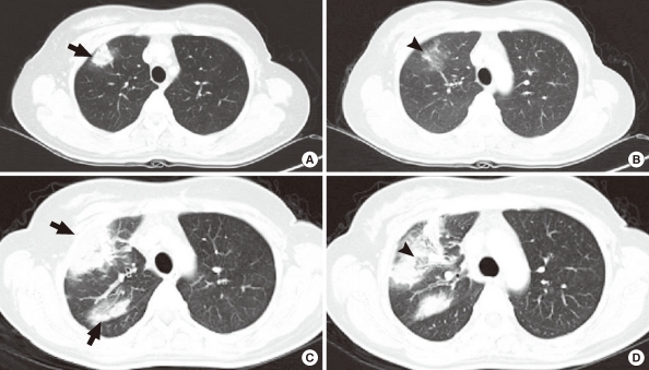
Figure 2.
(A, B) Chest computed tomography (CT) scan at the emergency room. Patchy consolidative mass-like lesion in the right upper lobe (RUL) (arrow) with focal ground glass opacity (GGO) (arrowhead). (C, D) After 1 week of empirical antibiotics for community acquired pneumonia, RUL consolidation and consolidation near the interlobar fissure and hilum increased in extent (arrows), and GGO of the RUL increased in extent (arrowhead).

