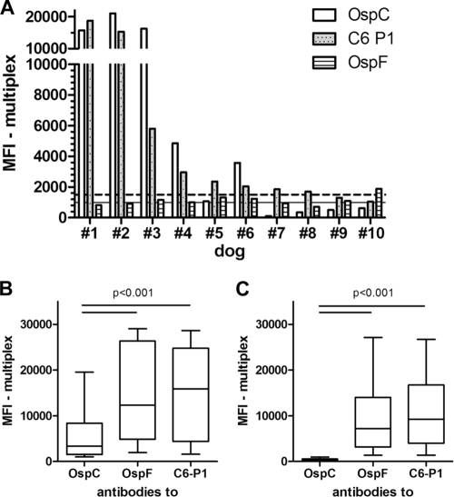
Fig 3.
Comparison of antibodies to OspC, OspF, and C6 obtained from canine patient sera. Antibodies were determined by multiplex analysis. Canine patient sera were submitted to the Animal Health Diagnostic Center at Cornell University between July 2008 and January 2009. (A) OspC antibody values of nine dogs (#1 to 9) with a C6+/OspF− and one dog (#10) with C6−/OspF+ detection pattern in serum. The antibodies to OspC, OspF, and C6 P1 were analyzed in the multiplex assay. These 10 samples, with disagreement on the Lyme antibody status interpretation based on C6 and OspF, were obtained from a total of 125 canine patient sera. For the remaining 115 samples, the multiplex assay interpretation based on OspF and C6 was in agreement. The C6 values decrease from dog 1 to 10. The horizontal lines show the positive OspC (intact line) or C6 and OspF (dotted line) cutoff values. (B) Sera from 21 dogs with antibodies to all three infection markers. (C) Sera from 43 dogs with antibodies to OspF and C6 that were negative for antibodies to OspC. The horizontal lines in plots B and C indicate significant differences between the antigens. The P value applies to both lines in each plot.

