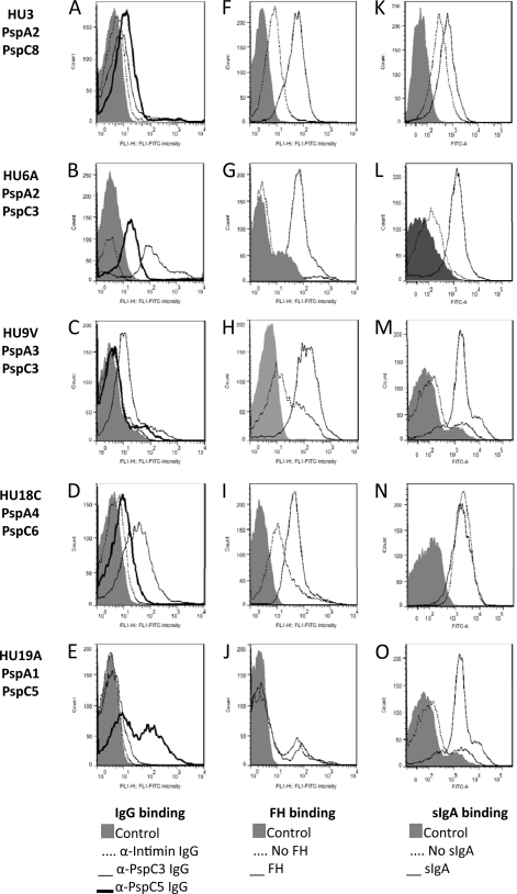
Fig 4.
Flow cytometry analysis of binding of IgG, FH and sIgA to clinical isolates. Binding of IgG (A to E), FH (F to J), and sIgA (K to O) to intact bacteria was analyzed by flow cytometry. Pneumococcal isolates HU3 (A, F, and K), HU6A (B, G, and L), HU9V (C, H, and M), HU18C (D, I, and N), and HU19A (E, J, and O) were used. Bacteria incubated only with the detection antibody conjugated with FITC were used as a control for each strain. Results for samples without incubation with FH and sIgA are also shown.

