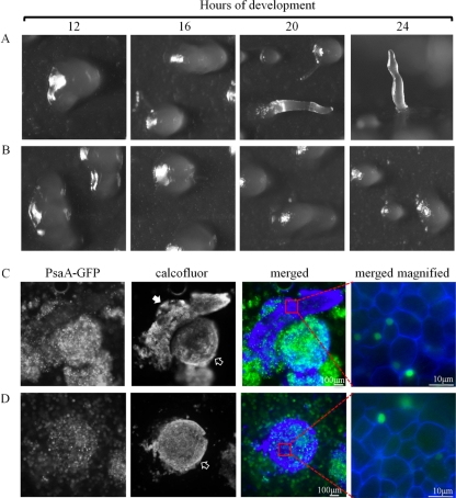
Fig 14.
The effect of BME on development of PsaA-GFP-overexpressing cells. A developmental sequence of PsaA-GFP-expressing cells treated with 600 (A) or 900 (B) μM BME and allowed to develop on filters for up to 24 h. The 24-h image in panel A is a side view; the rest of the images are top views. (C and D) Calcofluor-stained cells of the structures formed on 600 (C) and 900 (D) μM BME at 24 h. These correspond to structures seen at 24 h in panels A and B, respectively. Solid arrow points to finger, and empty arrows point to mounds. The last images in panels C and D show the stalk cells that comprise these structures. This experiment was independently replicated 4 times. At least 10 finger- and mound-like structures were stained and viewed each time.

