Fig 3.
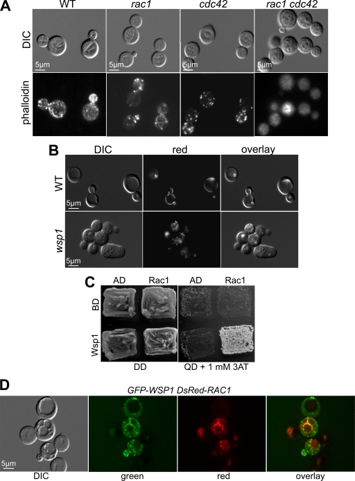
Wsp1 is an effector of Rac1 in actin organization. (A) F-actin was visualized using rhodamine-conjugated phalloidin staining of the indicated strains. WT, wild type. DIC, differential interference contrast. (B) Rac1 is enriched in the vacuolar membrane. The DsRed-Rac1 fusion protein was expressed in the wild-type and wsp1 mutant strains. Rac1 localization near the vacuolar membrane in the wild-type strain was disrupted in the wsp1 mutant. (C) Rac1 interacts with Wsp1 in a yeast two-hybrid assay. The two-hybrid yeast strain was incubated for 3 days on either DD (double-dropout medium [SD-Leu-Trp]), indicating the presence of both vectors, or QD (quadruple-dropout medium [SD-Leu-Trp-His-Ade]), suggesting interaction. AD, empty activation domain vector; BD, empty binding domain vector; Rac1, C. neoformans RAC1 allele in BD; Wsp1, C. neoformans WSP1 allele in AD. (D) GFP-Wsp1 colocalizes with DsRed-Rac1. The GFP-Wsp1 and DsRed-Cdc42 fusion proteins were coexpressed in the wsp1 rac1 double mutant strain. Colocalization of the fluorescent signals around the vacuole was visualized in the overlay image.

