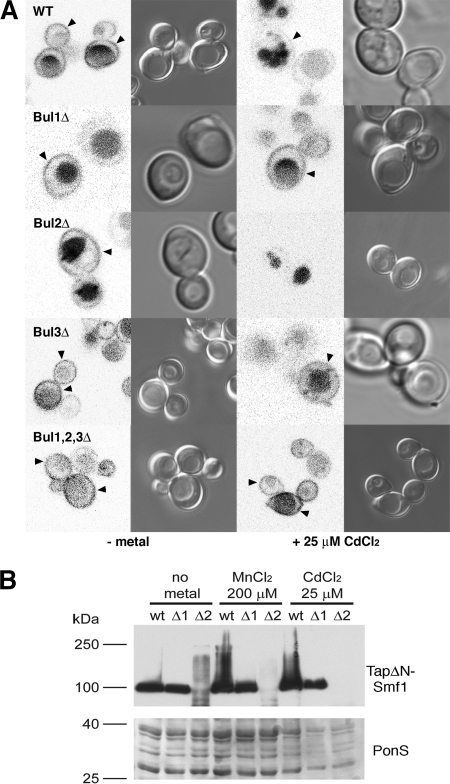
Fig 3.
Sorting of mCherryΔNSmf1p in metal-depleted medium following the addition of CdCl2. (A) mCherry fluorescence and differential interference contrast microscopy images of wild-type and single BUL gene deletion strains expressing mCherryΔNSmf1p following growth on metal-depleted media (−metal) and 2 h after the addition of 25 μM CdCl2. The arrowheads indicate plasma membrane localization. (B) (Top) Immunoblots against Tap-tagged ΔNSmf1p in total protein extracts from wild-type and bul1Δ (Δ1) and bul2Δ (Δ2) strains. (Bottom) PonceauS staining was used as a loading control.

