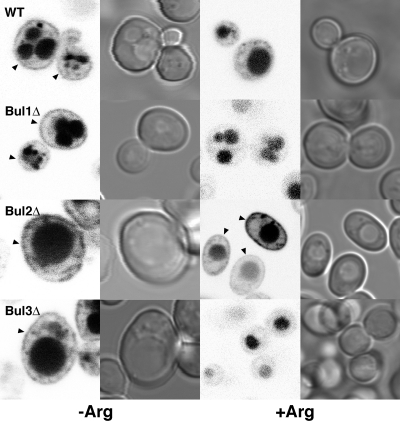
Fig 5.
Sorting of GFPCan1p following growth in excess arginine. Shown are GFP fluorescence and differential interference contrast microscopy images of wild-type and single BUL gene deletion strains expressing GFPCan1p. The images were obtained before (−Arg) and 2 h after (+Arg) the addition of 100 mg/liter arginine to log-phase cells grown in arginine-free synthetic complete medium. The arrowheads indicate plasma membrane localization.

