Fig 5.
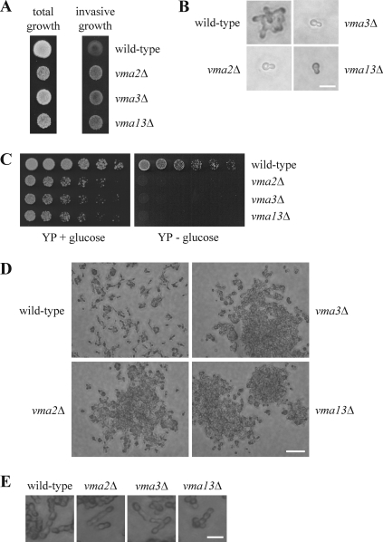
Cells lacking V-ATPase activity are hyperinvasive. (A) Deletion of V-ATPase subunits leads to stronger agar invasion. Cells (104) of the indicated strains were spotted on a YPD plate and grown for 3 days at 30°C. Photographs were taken before (total growth) and after (invasive growth) rinsing with water. Note that agar invasion by the vma mutants is markedly stronger than that by the wild type even though these strains have a greatly reduced growth rate. (B) vma mutants fail to form colonies in the absence of glucose. Cells (104) of the indicated strains were spread on SC medium lacking glucose and were incubated for 18 h at 30°C. Bar, 10 μm. (C) Cells lacking V-ATPase activity are unable to grow on YP medium lacking glucose. Serial dilutions (1:3) of the indicated strains were spotted on YP plates with or without glucose and incubated for 2 days at 30°C. (D) vma mutants form cell clusters in response to nutrient deprivation. Cells (103) of the indicated strains were spotted on a YPD plate and grown for 3 days at 30°C. Cells that did not invade the agar were washed off with water. Bar, 50 μm. (E) Morphology of cells grown on agar. The indicated strains were treated as described for panel D but incubated for 3 days at 30°C. Bar, 10 μm.

