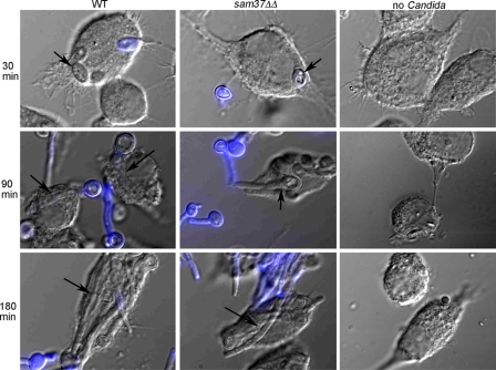
Fig 4.
Filamentation by the sam37ΔΔ mutant upon phagocytosis by macrophages. Figures are overlaps of DIC images and fluorescent images and were constructed using ImageJ. Calcofluor white staining was used to differentiate intracellular from external C. albicans (the yeast cells inside the macrophage do not stain with calcofluor white). Internalized C. albicans cells are marked by the arrows.

