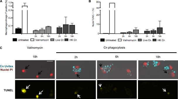
Fig 6.
DNA fragmentation and membrane permeability in J774.16 macrophages. J774.16 macrophages did not manifest increased membrane permeability or DNA fragmentation after phagocytosis of C. neoformans. (A and B) Quantification of the number of cells positive for membrane permeability (A) or DNA fragmentation (B). Treatment with valinomycin at 400 nM for 18 h was used as a positive control, while uninfected macrophages were used as the negative control. (C) Representative images of macrophages positive for TUNEL staining after treatment with 400 nM valinomycin (positive control) or after infection with live C. neoformans (data for HK C. neoformans are not shown). Nuclei positive for DNA fragmentation have apoptotic cell morphology and are rapidly engulfed by a neighboring cell (arrows). Scale bar, 20 μm. Experiments were performed in duplicate wells for DNA fragmentation or in triplicate wells for membrane permeability measurements. Experiments were performed twice in J774.16 macrophages, and the results of one experiment are shown. Experiments were performed once for BMDM, and same results as observed for J774.16 were obtained.

